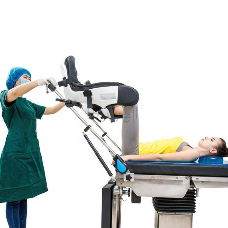
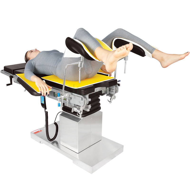
Lithotomy position, one of the surgical positions. It is characterized by the patient lying on his back, with his legs placed on the leg frame and his buttocks moved to the bedside, which can expose the perineum to the maximum extent. It is mostly used in anorectal surgery and gynecological surgery.
The lithotomy position is often used in vaginal hysterectomy, rectal surgery, cystoscopy and other operations. Specific placement method is: the patient supine, put on leg cover. The client is then lowered so that the sacral tail is slightly beyond the lower edge of the backplane and the legs are placed on the leg rack.
Common adverse reactions include local skin compression, peroneal nerve injury and venous thrombosis.
Causes of adverse reactions
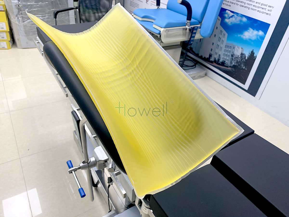
Local skin compression: The patient already had blood circulation disorder in lower limbs before surgery; Bone carina without proper gel pad; Uneven gel pad; The pressure of the surgeon.
Vein thrombosis: the popliteal artery and popliteal vein are the main blood vessels maintaining the blood circulation in the lower leg. The popliteal artery and popliteal vein lack the protection of muscle and adipose tissue at the popliteal fossa. Therefore, long-term compression of the popliteal fossa will cause blood circulation disorder in the lower leg, resulting in vascular intima injury or venous thrombosis.
The factors causing excessive popliteal fossa compression are: the constraint band is too tight, improper position; The knee bend Angle is too small.
A new method of positioning the lithotomy position , patients with upper limbs to own sheets on both sides of the fixed to the body, can avoid brachial plexus upper limb outreach lead to damage to the leg holder frame hold patients crus, gel pads are placed in the leg, leg of gel pad should be the first to spin direction in packages, lumbosacral region soft mat mat thickness about 10 cm high, make the operation fully exposed, convenient operation, the waist dangling in thin soft mat mat, can reduce stress, prevent total nerve Lesions and sacral skin lesions.
Injury of the common peroneal nerve The common peroneal nerve is a branch of the sciatic nerve that goes around the fibula neck and crosses the peroneal longus muscle to the front of the calf. The common peroneal nerve bypassed the fibula neck near the skin and lacked the protection of muscle-adipose tissue. Prolonged compression at this point can result in damage to the common peroneal nerve.
